Aim : to guess how an image would look like if it had been taken by another imaging tool Application : to convert a human body scan obtained by Magnetic Resonance (MR) into a scan which whould have been obtained by Computed Tomography (CT), which are two kinds of medical scanners based on completely different physics and technology Difficulties : no trivial correspondence between intensities of MR and CT scans. In MR scans, air and bone are black, fat is white, and brain tissue is grey; whereas in CT scans, air is black, bone is white, fat and brain tissue are grey. So that something black in a MR scan could be black (air) or white (bone) in CT scans.
Method : to learn locally the correlation between intensities of both scans, in a training set of pairs of corresponding MR and CT scans
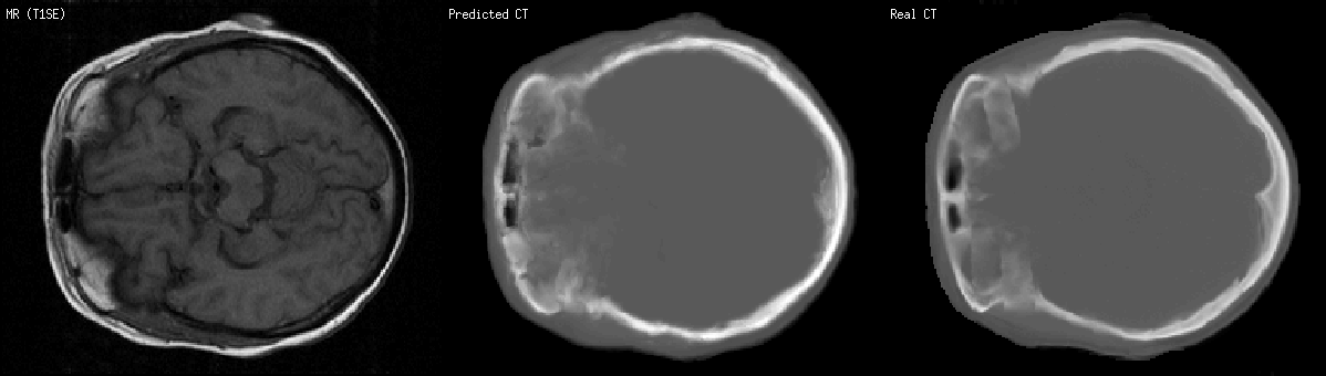
Example : a MR scan slice of a human brain, its predicted CT scan slice computed based on a training set of other MR and CT scans, and the real CT scan (i.e. the ground truth in order to see if predictions are good):
The two black holes in all images stand for cavities (so, air) in the forehead. White in the CT images stand for the skull. In the MR image, they all look black, so one cannot distinguish bone from air in MR scans just from the pixel intensity. Image patches and location are considered, in order to bring more information.
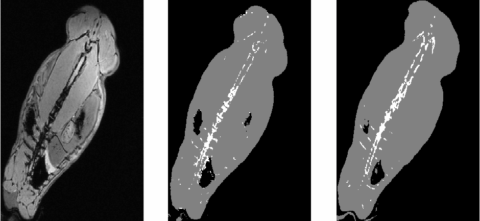
Another example, with slices of scans of a rabbit: In the CT images, the rabbit spine appears in white. The real CT scan does not correspond exactly to the MR scan because the rabbit was not set exactly in the same posture in the two scanners.
Guillaume Charpiat, Matthias Hofmann and Bernhard Schölkopf, Chapter Kernel methods in medical imaging, chapter of the book Biomedical Image Analysis: Methodologies and Applications, N. Paragios, J. Duncan & N. Ayache Editors, Springer, 2008. [bibtex]
Matthias Hofmann, Florian Steinke, Verena Scheel, Guillaume Charpiat, Mike Brady, Bernhard Schölkopf and Bernd Pichler, MR-Based PET Attenuation Correction: Method and Validation, IEEE Nuclear Science Symposium and Medical Imaging ConferenceNSS-MIC 2007. [bibtex]